
Glaucoma is a type of eye disease where the pressure within the eye is greatly increased. The high pressure results in a buildup of fluid within the eye and over time, lead to a damaged optic nerve. As it is evident, glaucoma is a serious condition and is best treated immediately and at the earliest possible stage. Various general eye tests also test for glaucoma on a regular basis but once identified, you will have to obtain the necessary treatment to preserve your eyesight. Glaucoma is diagnosed using various tests and tools:
The Tonometer – This device is designed to measure eye pressure. At first, an eye drop is placed in your eye and it is examined with a slit lamp by your doctor. This provides the doctor with a magnified view of your eye. The tonometer is a plastic prism that is moved forward until it barely touches the cornea. This test is easy and has no discomforting effects to the patient.
The Pachymeter – The pachymeter is used to measure the thickness of the central cornea. Your eyes will first be anesthetized to allow a small probe to be placed perpendicular to the center of the cornea. The central corneal thickness is an essential measurement to determine the levels of intraocular pressure.
The Visual Field Test – Elevated levels of intraocular pressure leads to optic nerve damage. The visual field test can help determine the amount of damage. This test allows the doctor to figure out if you have lost your peripheral vision. The test is conducted in several different ways.
Ophthalmoscopy – The Ophthalmoscope is a device that allows your doctor to see directly into the optic nerve through the pupil. The color and appearance of the nerve will tell the doctor whether it is damaged from glaucoma or not. The device also allows the doctor to get a measure of the extent of damage to the optic nerve. Ophthalmoscopy tests are essential in monitoring and diagnosing glaucoma.
Imaging – Imaging is done with the aid of various sophisticated systems that are available to analyze the retinal nerve fiber layer and the optic nerve which are susceptible for glaucoma damage. The devices used for imaging include laser polarimetry, laser tomography and ocular coherence tomography. They are designed to provide doctors with quantitative measures of the structured within the eye.
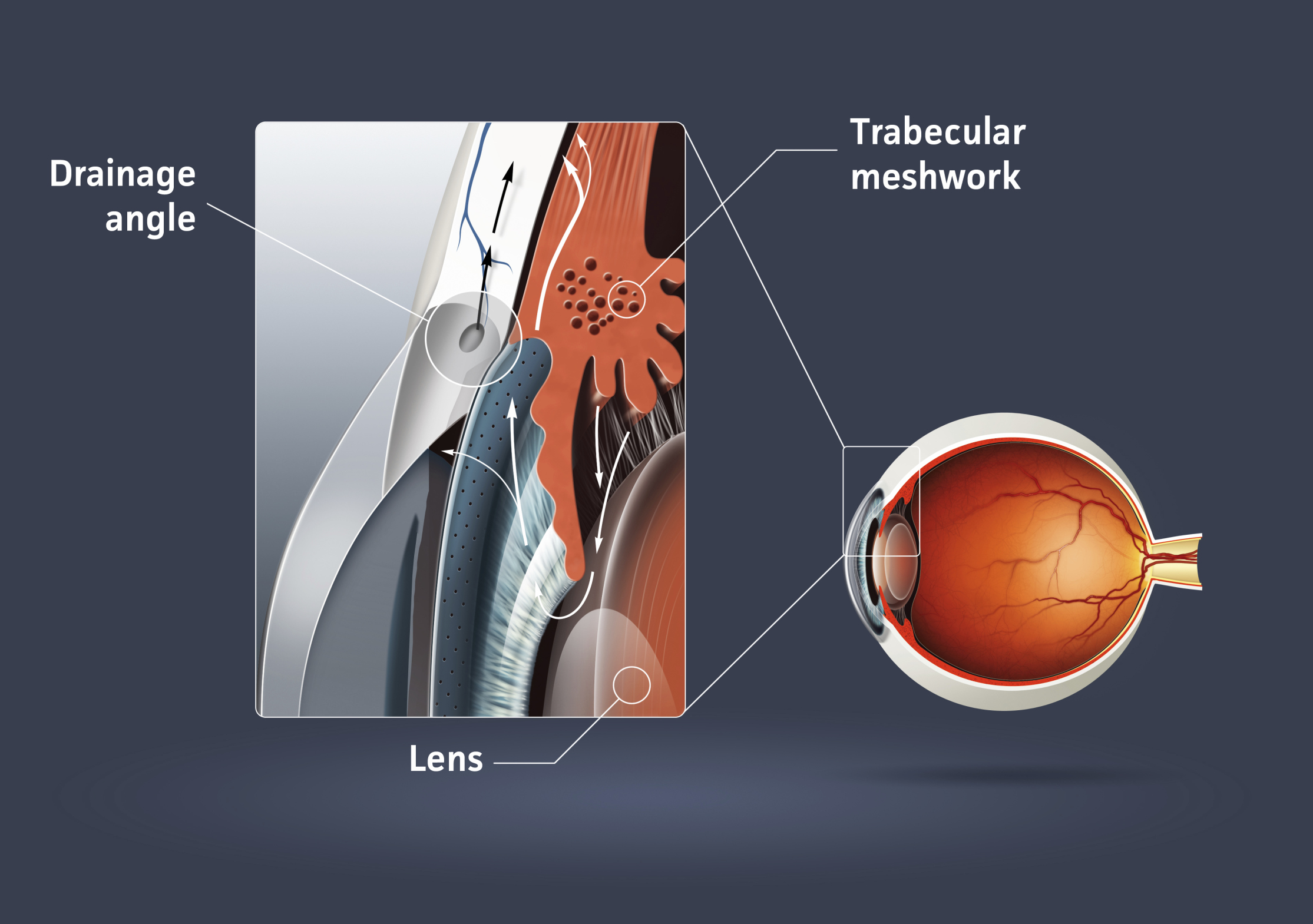
Gonioscopy – A gonioscopy may be required if your doctor needs to examine the trabecular meshwork in the eye as well as the angle in which the fluid drains from the eye. The eye will be numbed before the doctor places a contact lens, fitted with mirrors to view the inside of the eye. The doctor can use the lens to determine the angle as well as to view the interior of the eye.