Illustrated sliced eyeball retina diagram
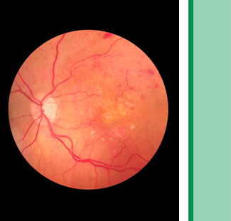
A thin, membranous lining, the retina is located at the back of the eye. It serves an important function: sending visual messages through the optic nerve to the brain.

During a retinal exam, Dr. Monroe Benaim (our ophthalmologist) will dilate your eyes, in order to take a closer look at the back of your eye. Then, he’ll use a special magnifying lens, which allows him to look for common retina problems.

The sooner a retina condition is caught, the sooner it can be treated. Book an appointment today to protect your vision or read on to learn about common retina problems and treatment.

Book an appointment

Diabetic Retinopathy

Diabetic retinopathy is the most common eye condition associated with diabetes. If you have diabetes, your body can’t regulate blood sugar levels correctly, and high blood sugar levels can cause damage to the blood vessels in your body, especially in the retina. Damage to the retina, known as diabetic retinopathy, can ultimately cause vision loss. Patients who are able to control their diabetes reduce their chance of diabetic retinopathy. In minor cases, laser treatment is used to seal blood vessels. In more severe cases, the vitreous gel may have to be surgically removed.

You can significantly reduce your risk of developing diabetic retinopathy by:

  • Monitoring blood pressure to keep it under control
  • Seeking appropriate care
  • Maintaining a healthy diet
  • Exercising regularly
  • Visiting an ophthalmologist for a yearly dilated eye exam

There are typically no symptoms in the early stages of diabetic retinopathy, so your vision may not be affected until the disease becomes severe. More than a third of patients diagnosed with diabetes do not receive the recommended vision care, and as a result, may be at risk for blindness.

It is important to pay close attention to changes in your vision. If your vision becomes blurred, spotty, or hazy, contact Dr. Benaim for a diabetic eye exam. Diabetic patients should get a comprehensive eye examination every year.

Floaters and Flashes

Floaters can appear in your vision as a variety of different shapes. They are often noticed when looking at a plain background, like a wall, or the sky. Floaters are caused by the clumping of the vitreous gel, which makes shadows on the retina, resulting in floaters in your vision.

A tear in the retina can cause floaters, a very serious problem. If a retinal tear is not treated then the retina may become detached from the back of the eye, and the only treatment for a detached retina is surgery. Other floaters are harmless and will disappear over time without the need for treatment. If you have suffered from floaters before, you should continue to see an eye doctor, who can examine you for any new ones that suddenly develop.

Dr. Benaim may dilate your pupils in order to conduct a full exam. This painless exam will allow your eye doctor to examine all the areas of your eye, including the retinal and vitreous gel.

Floaters are more common when people reach middle age. The vitreous gel becomes thicker or shrinks as we age, and this will form strands or clumps within the eye. The vitreous gel can begin to pull away from the back of the eye causing a posterior vitreous detachment, a common cause of floaters.

Flashes

Flashes are another common retina problem, especially as your eyes age. They can appear as flashing lights or lightening streaks.

Some people who experience the flashes of light see jagged lines in both eyes at once. These flashes last 10 to 20 minutes and are usually caused by a spasm of blood vessels in the brain, which is a migraine.

As you grow older, you might experience flashes more frequently. They can appear off and on for several weeks or months. If you notice the sudden appearance of light flashes, you should contact your eye doctor immediately, in case the retina has been torn.

Retinal Detachment

An animation of retinal detachmentIf the retina separates from the supporting structures in the back of the eye, it’s called retinal detachment. This retina condition is often caused by small holes formed on the retina. The eye fluid can enter the holes and start to peel the retina off the back of the eye. The holes can form as a result of trauma, retina degeneration, or after eye surgery.

The symptoms of retinal detachment may include the sudden development of floaters and flashes. Bleeding from the retinal tear can cause the floaters, and pull from the vitreous jelly, causing the flashes. If the retina starts to detach itself, a patient may see a blurry image in the edge of their vision. As the detachment progresses, the blurriness may enter the center. If the retina fully detaches, the macula will also detach and vision will become increasingly worse. Patients who experience these symptoms should see Dr. Benaim immediately.

If the retina has only experienced small tears, then treatment with a laser can usually fix the tear. Close monitoring will follow this treatment. If the retina becomes fully detached, a silicone band (called a scleral buckle) may be placed behind the eyelids to help reattach the retina. A vitrectomy, a surgical procedure where the vitreous gel is removed, may be necessary to help repair the retina. If the retinal detachment involves only the peripheral retina, then surgical repair usually helps maintain good vision.

There is a possibility that some vision may still be lost, even after surgical reattachment of the retina, especially if the detachment has progressed enough to involve the macula, which is the center of vision.

To catch retina conditions and common retina problems early on, schedule an appointment with Dr. Benaim at 561-747-7777.